Abstract
Introduction:
The use of an umbilical catheterization is a usual practice in neonatal units. The insertion of the catheter has potential complications.Case Presentation:
Here, we report on our observation of a seven-day-old female newborn admitted for an abdominal distention and vomiting bile. Initially, diagnosis was midgut volvulus, for which an operation was performed. During the surgery, no intestinal malrotation, mesenteric defect or atresia was observed. Postoperative diagnosis was abdominal wall hematoma and rand ligament and ileus, as well as, sub-capsular liver hematoma. The patient had been hospitalized at birth at a neonatal intensive care unit (NICU). With the appearance of icterus on the first day of life, at the NICU tried to insert the umbilical catheter that had been filed.Conclusions:
The complication found in the patient was the result of an aggressive act (the umbilical catheter insertion). This intervention should not be carried out unless there are clear indications, and if so, it should be done with much care.Keywords
1. Introduction
Use of umbilical catheter is a common procedure at neonatal intensive care units (NICUs) that is used for replacement transfusion in the treatment of erythroblastosis fetalis. In addition, umbilical catheter has been used for drawing blood samples, measuring venous blood pressure, and administering fluids, nutrition and medications, blood products, and exchange therapies in sick neonates (1). Use of umbilical catheters has several complications as reported by previous studies, such as thrombosis, embolization, hemorrhage, abscess, arrhythmias, effusions, portal hypertension, renal vein perforation, necrotizing enterocolitis and perforation of the colon, sepsis, cardiac tamponade, and ischemic injury of the extremities (1-6).
Liver hematoma is a rare but life-threatening complication of umbilical catheterization that is a result of direct injury through liver parenchyma. Here, we report on a newborn with liver hematoma as a rare and serious complication duo to unsuccessful of umbilical catheterization during previous hospitalization.
2. Case Presentation
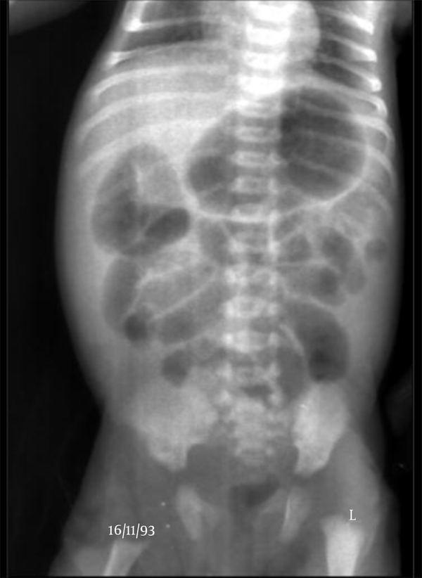
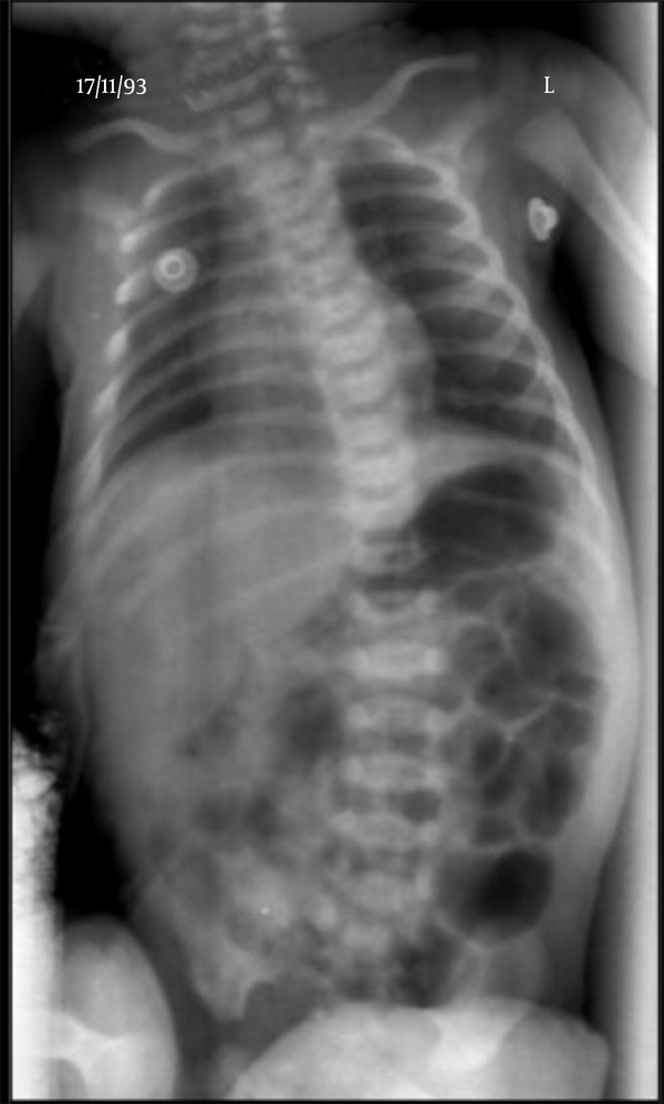
The patient was a seven-day-old girl born at 37 weeks of gestation by normal vaginal delivery. Her birth weight was 2723 g, and now she was 2520 g. She had B+ blood type and was the first child of a mother, who had B-blood type. The patient had no particular problems at birth, breast-feeding had been tolerated, and meconium passed during the first day of birth. The abdomen was markedly distended, vomiting bile, and bilirubin level was 17, therefore, she was admitted to Sheikh children’s hospital. Six days before hand, the patient was referred to a medical center for treatment of icterus and had unsuccessful umbilical venues catheter and was then discharged. After 20 hours, the patient with symptoms of vomiting and abdominal distention was referred to our center. She was transferred to the neonatal intensive care unit (NICU) due to her critical condition. After admission, routine laboratory examinations were performed. Her rectal examination was normal. Initial laboratory values included a white blood cell count of 12000/mm3, a platelet count of 329000/mm3 and prothrombin time of 14 seconds. Blood electrolyte level from the umbilical artery was Na 134 mg/dL, K 4.5 mg/dL. Because of the urgency of the situation, intestinal obstruction or colon volvulos was diagnosed only with X-ray and according to the sign and physical examination and abdominal graphs, the patient with suspicion based obstruction, was scheduled for surgery for midgut volvulus. There was no intestinal malrotation, mesenteric defect or atresia. Postoperative diagnosis was abdominal wall hematoma and rand ligament and ileus, as well as, sub-capsular liver hematoma. Two days after the operation, abdominal ultrasound and upper gastrointestinal (GI) series were performed, with completely normal results. The patient’s history was obtained from her parents and they noted when she was admitted to the hospital for icterus she had no IV and had tried several times to umbilical catheter that has failed. After the surgery and evacuation of liver hematoma and flushing her stomach with good tolerance of milk, she was discharged. (Figure 1 - 2, Table 1).
Preoperative Radiograph of the Abdomen

Postoperative Radiograph of the Abdomen

The Laboratory Findings
| Test | First Results |
|---|---|
| Blood | |
| Sugar (mg/dl) | 166 |
| Creatinine (mg/dL) | 0.5 |
| Na (mEa/L) | 134 |
| K (mEa/L) | 4.5 |
| Ca (mg/dL) | 9 |
| P (mEa/L) | 6 |
| Total bilirubin | 9.1 |
| ALP | 430 |
| PT/PTT | 14/34 |
| ESR | 13 |
| CRP | 45 |
| CBC | |
| WBC (cells/μL) | 12000 |
| RBC (cells/μL) | 4000000 |
| Hb (gr/dL) | 13 |
| Hct | 39 |
| Plt (mL) | 329000 |
| MCV(fl) | 99 |
| MCH | 33 |
| MCHC | 33 |
3. Discussion
Complications of umbilical vein catheterization include vascular injury, thrombosis, infection, hepatic necrosis and portal hypertension (7). Hepatic injures are more severe than other complications with a high risk for morbidity and mortality (8). Lim-Dunham et al. (9) studied the characteristic sonographic findings of hepatic erosion by UVCs. They showed that all neonates had abdominal distension within nine days of catheter placement, and in all of them, the UVC tip was located below the hemidiaphragm and superimposed over the liver. Result of sonographic examination revealed intraparenchymal liver lesions with an echogenic rim and a hypoechoic center. In these patients, treatment consisted of removing the UVC. Resolution of the parenchymal lesion occurred within two to 18 months. These findings demonstrate that, in case of abdominal distension, a high index of suspicion of UVC erosion into the liver should be taken into consideration. A UVC is routinely inserted and advanced from the umbilicus, blindly, to a predetermined length. The reported rates for catheter misplacement range from 20% to 37% (10). In most cases, positioning of the UVC is confirmed by radiography, which results in excessive radiation exposure, particularly in critically ill infants. However, some studies such as that of Yigiter have shown that radiography is unreliable in determining exact catheter placement (4).
On the other hand, hepatic abnormalities may occur due to mal-position of umbilical catheterization in infants, and this has been associated with high rates of mortality varying from 50% to 75% (11). The catheter must be placed in a high position, which is defined as its tip in the inferior vena cava, just before entrance to the right atrium. In this position, the tip should be seen above the diaphragma in the abdominal radiograph, at the level of the eighth and ninth thoracic vertebrae (12). The principal indication for umbilical vein catheterization is to gain vascular access during emergency resuscitation. Venous catheter access was required in all very preterm babies and any other baby requiring respiratory support, for urgent vascular access in resuscitation for administration of adrenaline and volume expansion or exchange transfusion. In addition, was needed infusion of hypertonic solutions for example in resistant hypoglycaemia requiring more than 10% dextrose or TPN (13). It should be noted that absolute contraindications to umbilical catheterization include omphalitis, peritonitis and necrotizing enterocolitis.
Umbilical catheterization that is used commonly in NICU has led to an increased incidence of adverse effects. Impress hepatic by umbilical catheterization has been reported in other studies and mentioned as a rare but serious complication of umbilical catheterization in infants (14). Different methods, such as X-ray, ultrasonography, echocardiography and electrocardiography, have been mentioned in the literature for verifying the correct positioning of umbilical catheterization. Because of difficulties in deciding the correct position with X-ray, ultrasonography evaluation seems to be a more valuable technique to diminish complications with high mortality (15).
To the best of our knowledge, two reports of hepatic abscess by umbilical catheter have been published in the English literature to date (11, 15). All neonates had abdominal distension, and in all of them, the tips of the umbilical catheter were located below the diaphragm and superimposed over the liver on plain radiographs. Also, in all 11 neonates in the study of Lim-Dunham, the catheter tip was between T8 and T12, projecting over the liver. Lim-Dunham et al. emphasized on the importance of a high index of suspicion of hepatic erosion by the UVC in neonates with suboptimal placed catheters (9).
Our patient also had abdominal distension and vomiting bile. In addition, in previous hospitalization, she had unsuccessful placement of umbilical catheter for treatment of icterus. It is important that patients, who are candidates for umbilical catheterization, are in emergency situations, and have the required indications.
3.1. Conclusion
The complication in our patient was the result of an aggressive act (umbilical catheter investment) at the hospital. This intervention should not be carried out unless there are clear indications, and if so, it should be done with much care. Contrast material injection through the UVC for diagnosis and diagnostic paracentesis can be avoided if physicians take care of such neonates by being familiar with the clinical, radiologic, and sonographic findings associated with hepatic erosion. Furthermore, early diagnosis can prevent life-threatening complications caused by hepatic necrosis and hemorrhage.
Acknowledgements
References
-
1.
Green C, Yohannan MD. Umbilical arterial and venous catheters: placement, use, and complications. Neonatal network. 1998;17(6):23-8.
-
2.
Lam HS, Li AM, Chu WC, Yeung CK, Fok TF, Ng PC. Mal-positioned umbilical venous catheter causing liver abscess in a preterm infant. Biol Neonate. 2005;88(1):54-6. [PubMed ID: 15802908]. https://doi.org/10.1159/000084718.
-
3.
Al Nemri AM, Ignacio LC, Al Zamil FA, Al Jarallah AS. Rare but fatal complication of umbilical venous catheterization. Congenit Heart Dis. 2006;1(4):180-3. [PubMed ID: 18377544]. https://doi.org/10.1111/j.1747-0803.2006.00031.x.
-
4.
Yigiter M, Arda IS, Hicsonmez A. Hepatic laceration because of malpositioning of the umbilical vein catheter: case report and literature review. J Pediatr Surg. 2008;43(5):E39-41. [PubMed ID: 18485935]. https://doi.org/10.1016/j.jpedsurg.2008.01.018.
-
5.
Ancora G, Soffritti S, Faldella G. Diffuse and severe ischemic injury of the extremities: a complication of umbilical vein catheterization. Am J Perinatol. 2006;23(6):341-4. [PubMed ID: 16841280]. https://doi.org/10.1055/s-2006-946721.
-
6.
Hermansen MC, Hermansen MG. Intravascular catheter complications in the neonatal intensive care unit. Clinics in perinatology. 2005;32(1):141-56.
-
7.
Hui JY, Lo KK, Lo J, Chan ML, Chan JC. Neonatal total parenteral nutrition ascites secondary to umbilical venous catheterization. J Hong Kong Coll Radiol. 2001;4:288-90.
-
8.
Morag I, Epelman M, Daneman A, Moineddin R, Parvez B, Shechter T, et al. Portal vein thrombosis in the neonate: risk factors, course, and outcome. J Pediatr. 2006;148(6):735-9. [PubMed ID: 16769378]. https://doi.org/10.1016/j.jpeds.2006.01.051.
-
9.
Lim-Dunham JE, Vade A, Capitano HN, Muraskas J. Characteristic sonographic findings of hepatic erosion by umbilical vein catheters. J Ultrasound Med. 2007;26(5):661-6. [PubMed ID: 17460008].
-
10.
Tsui BC, Richards GJ, Van Aerde J. Umbilical vein catheterization under electrocardiogram guidance. Paediatr Anaesth. 2005;15(4):297-300. [PubMed ID: 15787920]. https://doi.org/10.1111/j.1460-9592.2005.01433.x.
-
11.
Tan NW, Sriram B, Tan-Kendrick AP, Rajadurai VS. Neonatal hepatic abscess in preterm infants:a rare entity? Ann Acad Med Singapore. 2005;34(9):558-64. [PubMed ID: 16284678].
-
12.
Detaille T, Pirotte T, Veyckemans F. Vascular access in the neonate. Best Pract Res Clin Anaesthesiol. 2010;24(3):403-18. [PubMed ID: 21033016].
-
13.
Symansky MR, Fox HA. Umbilical vessel catheterization: Indications, management, and evaluation of the technique. Pediatrics J. 1972;80(5):815-9. https://doi.org/10.1016/s0022-3476(72)80137-9.
-
14.
Mahajan V, Rahman A, Tarawneh A, Sant'anna GM. Liver fluid collection in neonates and its association with the use of a specific umbilical vein catheter: Report of five cases. Paediatr Child Health. 2011;16(1):13-5. [PubMed ID: 22211066].
-
15.
Bayhan C, Takci S, Ciftci TT, Yurdakok M. Sterile hepatic abscess due to umbilical venous catheterization. Turk J Pediatr. 2012;54(6):671-3. [PubMed ID: 23692799].